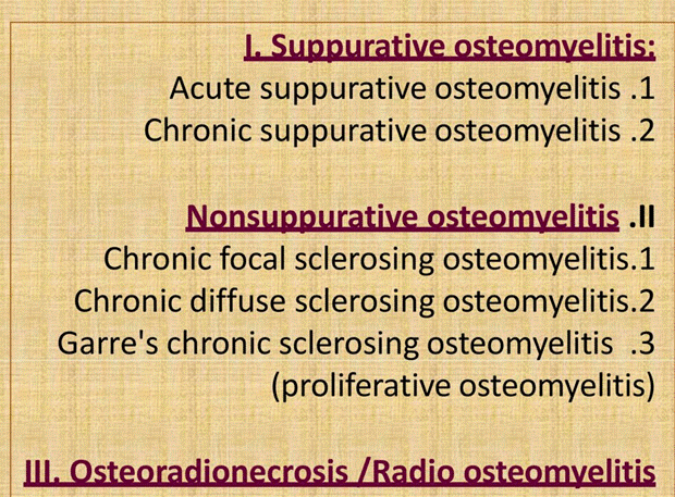
Osteomyelitis:It is an inflammation of the bone.

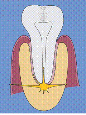
Potential spread of pus from a mandibular periapical abscess
Clinical features
Most patients with osteomyelitis are adult males, who have more dental infections than females. Almost all cases affect the mandible, which is less vascular than the maxilla.
Early complaints are severe, throbbing, deep-seated pain and swelling with external swelling due to inflammatory oedema.
Later, distension of the periosteum with pus and, finally, subperiosteal bone formation cause the swelling to become firm. The overlying gingiva and mucosa is red, swollen and tender.
Associated teeth are tender. They may become loose, and pus may exude from an open socket or gingival margins. Muscle oedema causes difficulty in opening the mouth and swallowing. Regional lymph nodes are enlarged and tender and anaesthesia or paraesthesia of the lower lip, caused by pressure on the inferior dental nerve, is characteristic.
Frequently, the patient remains surprisingly well but, in the acute phase, there may be fever and leucocytosis.

Radiographic changes do not appear until after at least 10 days, and radiographs can provide little useful information before this time except to identify a local predisposing cause. Later, there is loss of trabecular pattern and areas of radiolucency indicating bone destruction and sometimes widening of periodontal ligament. Affected areas have ill-defined margins and a moth-eaten appearance similar to a malignant neoplasm. Areas of dead bone appear as relatively dense areas which become more sharply defined as they are progressively separated as sequestra. Later, in young persons particularly, subperiosteal new bone formation causes a buccal swelling and appears as a thin, curved strip of new bone below the lower border of the jaw in lateral or panoramic radiographs.
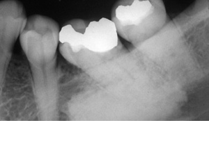
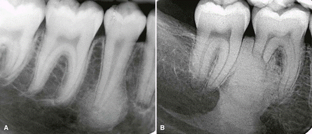
Osteomyelitis of the mandible following dental extractions. The outlines of the extraction sockets can be seen, together with dense sequestra of bone lying in a poorly circumscribed radiolucency.
Important predisposing conditions for osteomyelitis |
Local damage to or disease of the jaws • Radiation damage • Causes of osteosclerosis • Paget's disease • Fibro-osseous lesions, particularly cemento-osseous dysplasia • Osteopetrosis Impaired immune defences • Poorly controlled diabetes mellitus • Sickle cell anaemia • Chronic alcoholism or malnutrition • Drug abuse • Tobacco smoking • Malignant neoplasms and their treatment
|
Stages of development of the Disease progression
➢ The inflammatory process may spread through the bone to involve the marrow, cortex, cancellous portion, and periosteum.
➢ In the jaws, pyogenic organisms that reach the bone marrow from abscessed teeth or post surgical infection usually cause osteomyelitis.
➢ In some instances, no source of infection can be identified, and hematogenous spread is presumed to be the origin.
➢ In some patients, no infectious organisms can be identified, possibly because of previous antibiotic therapy or inadequate methods of bacterial isolation.
➢ Bacterial colonies also may be present in small, isolated pockets of bone that may be missed during sampling.
In patients with osteomyelitis, the bacteria and their products stimulate an inflammatory reaction in bone, causing resorption of the endosteal surface of the cortical bone. This resorption may progress through the cortical bone to the outer periosteum.
In young patients, in whom the periosteum is more loosely attached to the outer cortex of bone than it is in adults, the periosteum is lifted up by inflammatory exudate, and new bone is laid down
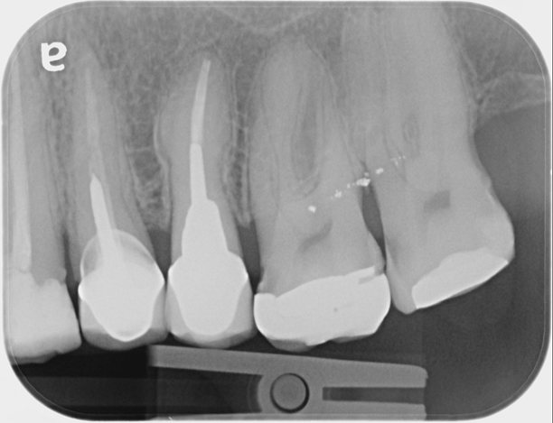
DBI (enostosis) in periapical positions. A, DBI around the apex of a second bicuspid. The periodontal membrane space is uniform in width. B, DBI associated with apical root resorption of a vital tooth. The most common site of DBI and root resorption is the mesial or distal root of mandibular first molars.
Chronic low-grade focal osteomyelitis and sclerosing osteitis
Summary of management of acute osteomyelitis |
|
Essential measures |
• Bacterial sampling and culture • Vigorous (empirical) antibiotic treatment • Drainage • Analgesics • Give specific antibiotics once culture and sensitivities are available • Debridement • Remove source of infection, if possible |
Adjunctive treatment
|
Sequestrectomy • Decortication if necessary • Resection and reconstruction for extensive bone destruction |
Acute osteomyelitis of the jaws: key features |
• Mandible mainly affected, usually in adult males • Infection of dental origin; anaerobes are important • Pain and swelling of jaw • Teeth in the area are tender: gingivae are red and swollen • Sometimes paraesthesia of the lip • Minimal systemic upset • After about 10 days, radiographs show moth-eaten pattern of bone destruction • Good response to prompt antibiotic treatment and debridement |
Chronic osteomyelitis
Chronic osteomyelitis is much more common than acute osteomyelitis and arises from infection by weakly virulent bacteria or in avascular bone. Most cases develop without a prior acute phase, and only rarely does acute osteomyelitis lead to chronic osteomyelitis. When it does, it usually follows inadequate treatment.
Like the acute condition, there are usually predisposing factors such as shown above.
However, local bone sclerosis or irradiation are factors much more likely to predispose to chronic osteomyelitis than acute.
Clinical features
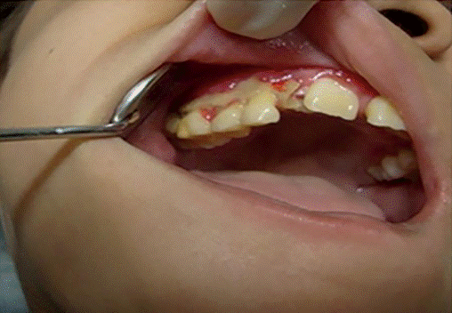
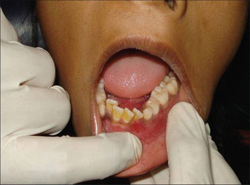
The picture is often dominated by persistent ache or pain, often relapsing, during a long period with a bad taste from pus draining to the mouth through sinuses. In more active phases there is swelling, increased pain and discharge, and increased tooth mobility. There may be exposed bone. Initially the original focus of infection can be identified, but chronic osteomyelitis may persist after its removal and the chronic infection becomes self-perpetuating in the bone.
Radiographic appearances are variable but sometimes distinctive with patchy and poorly defined radiolucency and sclerosis, sometimes resembling a malignant neoplasm. Sequestra may be identified, and there may be a periosteal new bone layer -proliferative periostitis
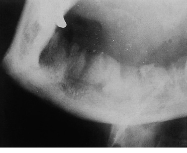
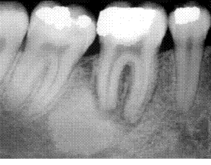
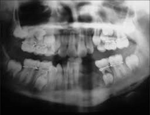
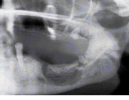
Chronic osteomyelitis. The extent of destruction is much more readily apparent than in acute osteomyelitis. Note the sequestra lying close to the lower border and the peripheral sclerosis. A slight convexity of subperiosteal new bone formation is evident below the lower border.
Chronic osteomyelitis with proliferative periostitis ( Garré’s osteomyelitis)
IT has a prominent periosteal inflammatory reaction as an additional component. It most often results from periapical abscess of a mandibular molar tooth, or from infection associated with tooth extraction or partially erupted molars. It is most common in children.
Clinical features
In the head and neck area, it is seen in the mandible. It typically involves the posterior mandible and usually is unilateral. Patients characteristically present with an asymptomatic bony, hard swelling with normal-appearing overlying skin and mucosa. On occasion, slight tenderness may be noted. This presentation necessitates differentiation of this process from benign mandibular neoplasms. Radiographs and a biopsy provide a definitive diagnosis.
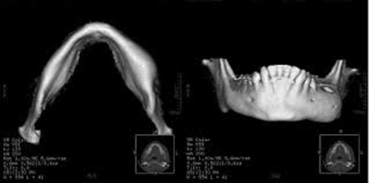
Radiographically, the lesion appears centrally as a mottled, predominantly lucent lesion in a pattern consistent with that of chronic osteomyelitis. The feature that provides the distinctive difference is the periosteal reaction. This, best viewed on an occlusal radiograph, appears as an expanded cortex, often with concentric or parallel opaque layers.Trabeculae perpendicular to the onion-skin layers may also be apparent.

Diffuse sclerosing osteomyelitis
Diffuse sclerosing osteomyelitis represents an inflammatory reaction in the mandible or maxilla, believed to occur in response to a microorganism of low virulence. Bacteria generally are suspected as causative agents, although they are seldom specifically identified. Chronic periodontal disease, which appears to provide a portal of entry for bacteria, is important in the origin and progression of diffuse sclerosing osteomyelitis. Carious nonvital teeth are less often implicated.
This condition may be seen at any age, in either sex, and in any race, but it tends to occur most often in middle-aged black women. The disease is typified by a protracted chronic course with acute exacerbations of pain, swelling, and occasionally drainage.
Radiographically, this process is diffuse, typically affecting a large part of the jaw.
The lesion is ill defined. Early lucent zones may appear in association with sclerotic masses. In advanced stages, sclerosis dominates the radiographic picture. Periosteal thickening may also be seen.
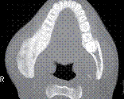
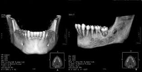
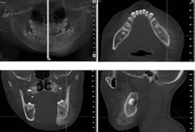
Diffuse sclerosing osteomyelitis of the right mandible in a computed tomography (CT) scan.
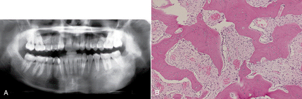
Diffuse sclerosing osteomyelitis of the left mandible. Biopsy specimen shows thick trabeculae, fibrous marrow, and scattered lymphocytes.
Osteoradionecrosis

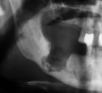
Left mandibular osteomyelitis with bone exposure (A) and sinus tract (B) following left mandibular radiotherapy in a patient receiving long-term bisphosphonate therapy. Panoramic dental X-ray shows mandibular lucencies (C). Note: The arrow shows mandibular lucencies.
Medication-related osteonecrosis of the jaws (MRONJ)
Drugs associated with jaw osteonecrosis |
• Antiresoptive • Bisphosphonates Alendronate Pamidronate Risendronate Zolendronate And other high potency types • Denosumab (antibody RANKL inhibitor) • Antiangiogenic |
Tyrosine kinase inhibitors Sorafenib Sunitinib • Bevacizumab (vascular endothelial growth factor inhibitor) • Rapamycin (mTOR [mechanistic target of rapamycin] inhibitor) |


Bisphosphonate-induced osteonecrosis. Non-healing extraction sockets in a breast cancer patient receiving bisphosphonate treatment.
Bisphosphonate-related osteonecrosis of the mandible.
Hypercementosis
Sclerotic bone islands