Periodontium - Healthy


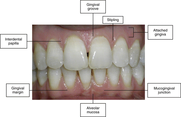
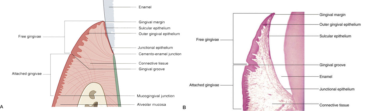
Diagram showing the anatomic landmarks of the gingiva
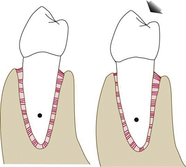
Left, Diagram of a mandibular premolar in a resting state.
Right, When a force is exerted on the tooth—in this case, in faciolingual direction (arrow)—the tooth rotates around the fulcrum or axis of rotation (black circle on root).
The periodontal ligament is compressed in areas of pressure and distended in areas of tension.

Section through a human jaw with a tooth in situ. The dotted line indicates the separation between the basal bone and the alveolar bone.

Relative proportions of cancellous bone and compact bone in a longitudinal faciolingual section of,
A, mandibular molars;
B, lateral incisors;
C, canines;
D, first premolars;
E, second premolars;
F, first molars;
G, second molars; and H, third molars.

The shape of the roots and the surrounding bone distribution in a transverse section of maxilla and mandible at the midroot level.
The variation of the position of the gingival margin with age.
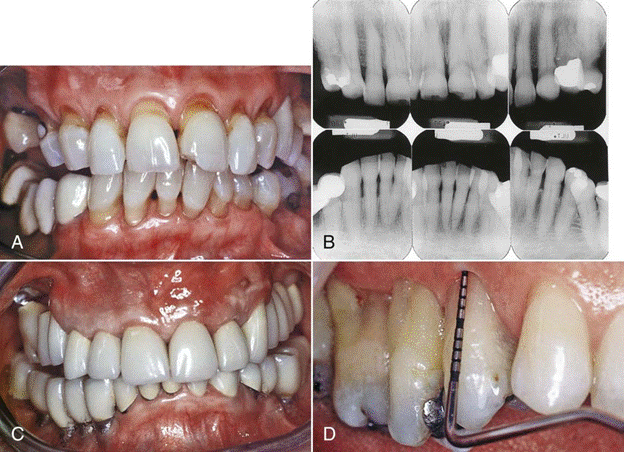
A, Overeruption with recession in an older individual (i.e., a 68-year-old woman) with generalized recession and a history of previously treated periodontitis