Cementoblastoma
Clinical & Radiographic features
Cementoblastoma, also known as true cementoma, is a rare benign neoplasm of cementoblasts that microscopically resembles an osteoblastoma but is connected or fused to the root of a tooth
It occurs predominantly in the second and third decades of life, typically before 25 years of age. There is no gender predilection.
It is seen more often in the mandible than in the maxilla and more often in posterior than in anterior regions.
It is intimately associated with the root of a tooth, and the tooth remains vital.
Cementoblastoma may cause cortical expansion and, occasionally, low-grade intermittent pain.
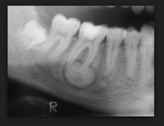
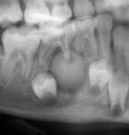
Radiographically, this neoplasm is an opaque lesion that fused rather than replaces the root of the tooth
It usually is surrounded by a thick uniform radiolucent ring that is contiguous with the periodontal ligament space and the advancing front of the tumor.


Cementoblastoma key features
Benign fibro-osseous/cementum jaw lesion
Young adults, mandible> maxilla
Attached to and replaces tooth root
Periodontal ligament space surrounds lesion
Opaque mass; may rarely cause cortical expansion
Histologic features of osteoblastoma
Treatment: Attached to tooth;tooth removed with full removal of lesion
No recurrence

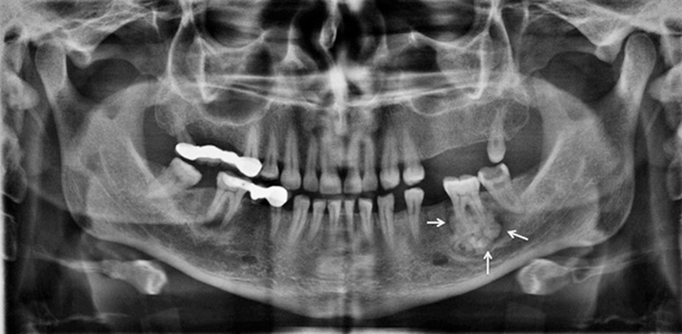
Cementoblastoma is seen at the periapex of the left second mandibular molar. A radiolucent halo is apparent at the periphery of the lesion (arrows).