Calcifying epithelial odontogenic tumor (Pindborg tumor)
Calcifying epithelial odontogenic tumor (CEOT), also known as Pindborg tumor, after the oral pathologist who first described the entity, is a benign tumor of odontogenic origin that shares many clinical features with ameloblastoma
Clinical features
CEOTs are seen in patients ranging in age from the second to the tenth decade, with a mean age of about 40 years. There is no gender predilection.
The mandible is affected twice as often as the maxilla, and a predilection for the molar-ramus region has been noted, although any site may be affected. Peripheral lesions, usually in the anterior gingiva, account for less than 5% of cases.
Jaw expansion or incidental observation on a routine radiographic survey is the usual way in which these lesions are discovered.
Radiographic features
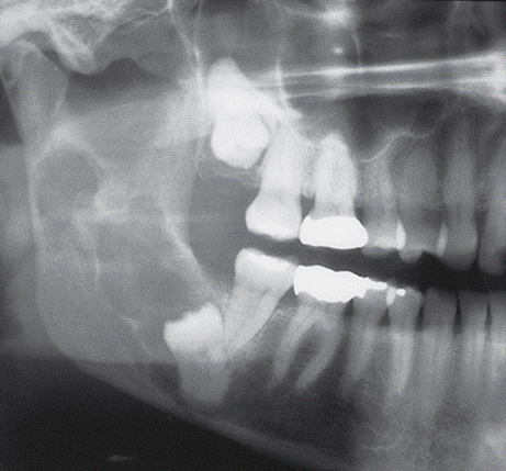
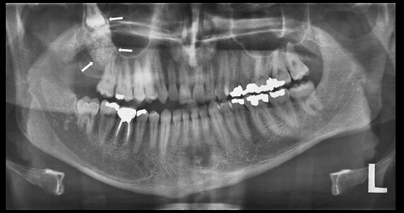
The lesions are often associated with impacted teeth.
The lesions may be unilocular or multilocular. Small loculations in some lesions have prompted use of the term honeycomb to describe this lucent pattern.
A CEOT may be completely radiolucent, or it may contain opaque foci, a reflection of the calcified amyloid seen microscopically.
The lesions are usually well circumscribed radiographically, although sclerotic margins may not always be evident.
Calcifying epithelial odontogenic tumour: key features |
Rare neoplasm of odontogenic epithelium Usually presents between ages 40 and 70 years Most commonly forms in posterior mandible Solid tumour, radiolucent, becoming a mixed radiolucency with time The only odontogenic tumour to contain amyloid Locally infiltrative like ameloblastoma Treated by excision with a small margin |

Calcifying epithelial odontogenic tumor. The multiloculated lesion extends from the third molar to the condyle.
Differential diagnosis
When this lesion is radiolucent, it must be separated clinically from dentigerous cyst, odontogenic keratocyst, ameloblastoma, and odontogenic myxoma.
Some benign nonodontogenic jaw tumors might also be considered, but these would be less likely, on the basis of age and location.
When a mixed radiolucent-radiopaque pattern is encountered, calcified odontogenic cyst should be considered in a clinical differential diagnosis. Other, less likely possibilities include adenomatoid odontogenic tumor, ameloblastic fibro-odontoma, ossifying fibroma, and osteoblastoma.

Treatment
This tumor has a locally infiltrative potential but apparently not to the same extent as ameloblastoma.
It is slow growing and causes morbidity through direct tumor extension. Various forms of surgery, ranging from enucleation to resection, have been used to treat CEOTs. The overall recurrence rate has been less than 20%, indicating that aggressive surgery is not indicated for the management of most of these benign neoplasms. Very rare examples of malignant transformation of this tumor have been reported and are associated with loss of p53 transcriptional activity.