Odontoma
Odontomas are commonest mixed odontogenic tumors, they are composed of both epithelial and mesenchymal dental hard tissues. They are developmental malformations (hamartomas).
These fully differentiated tissues are a composite of enamel and dentin. Biologically, odontomas can be regarded as hamartomas rather than neoplasms.
They are chance radiographic findings or present having prevented tooth eruption in children and adolescents.
In their early stages they are radiolucent, developing opaque flecks and then dense opaque masses as enamel and dentine form internally.
Occasionally they may erupt and then often become infected because of their convoluted shape and because no organised epithelial attachment forms.
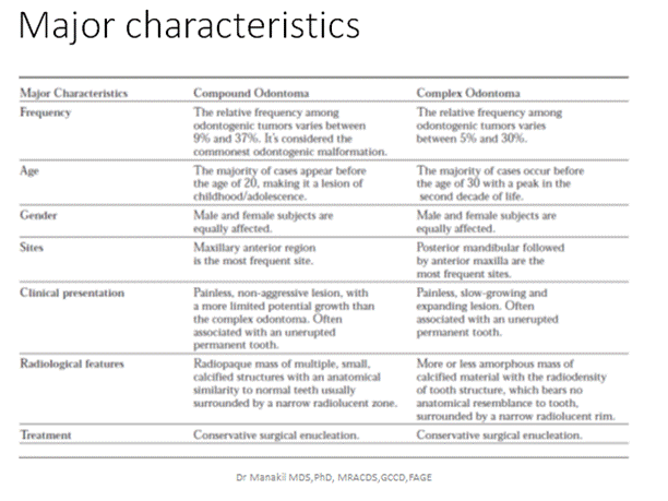
These calcified lesions take one of two general configurations. They may appear as numerous miniature or rudimentary teeth, in which case they are known as compound odontomas, or they may appear as amorphous conglomerations of hard tissue, in which case they are known as complex odontomas.
Clinical & radiographic features
Odontomas are lesions of children and young adults; most are discovered in the second decade of life . The range does, however, extend into later adulthood. There does not appear to be a significant gender predilection.
The maxilla is affected slightly more often than the mandible. There is also a tendency for compound odontomas to occur in the anterior jaws, and for complex odontomas to occur in the posterior jaws.
Clinical signs suggestive of an odontoma include a retained deciduous tooth, an impacted tooth, and alveolar swelling.
These lesions generally produce no symptoms
Radiographically, Lesions discovered during early stages of tumour development are primarily radiolucent, with focal areas of opacity representing early calcification of dentin and enamel. When calcification is complete, an irregular radiopaque mass is seen containing areas of densely radiopaque enamel
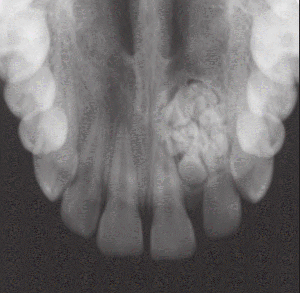
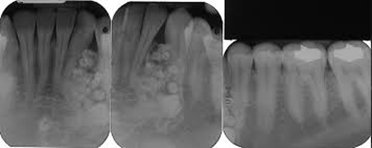
Compound odontome
These are clusters of many separate, small, tooth-like structures (denticles) within one crypt, the whole lesion usually no larger than 20 mm in diameter . This type is usually found in the anterior maxilla and causes minimal swelling.
Complex odontome
Complex odontomes consist of a single irregular mass as amorphous, opaque masses of hard and soft dental tissues, having no morphological resemblance to a tooth and frequently forming a cauliflower-shaped disorganised nodule of enamel and dentine. These may reach several centimetres in size and often expand the jaw.
Compound Odontoma
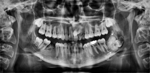
A complex odontoma with a radiolucent rim in conjunction with a corticated border around the lesion (arrows).
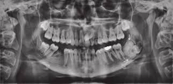
A panoramic view shows a large complex odontoma associated with the impacted left mandibular third molar.
Differential diagnosis
Compound odontomas are diagnostic on radiographic examination.
Complex odontomas usually present a typical radiographic appearance because of their solid opacification in relation to teeth.
The differential diagnosis might include other opaque jaw lesions such as focal sclerosing osteitis, osteoma, periapical cemental dysplasia, ossifying fibroma, and cementoblastoma.
Treatment
Odontomas have very limited growth potential, although an occasional complex odontoma may achieve considerable mass. Enucleation is curative, and recurrence is not a problem.
A rare variant known as odontoameloblastoma has been described. This is essentially an ameloblastoma in which there is focal differentiation into an odontoma. Until more is known of the behavior of this rare lesion, it should be treated as an ameloblastoma.
General characteristics of jaw lesions with a radiolucent rim
Radiolucent & Radiopaque or Mixed
