Effects on Surrounding Structures
The periapical inflammatory lesions may stimulate either the resorption of bone or the manufacture of new bone. The lamina dura around the apex of the tooth usually is lost. The sclerotic reaction of the cancellous bone may be limited to a small region around the tooth apex or may be extensive in some cases.
In rare instances in the mandible, the sclerotic reaction may extend to the inferior cortex. In chronic cases, external resorption of the apical region of the root may occur. If the lesion is long-standing, the pulp canal may appear wider than adjacent teeth. This is a result of the death of odontoblasts and subsequent cessation of the formation of secondary dentin.
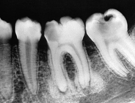
Periapical sclerosing osteitis associated with the first molar. This is called a sclerosing lesion because most of the lesion is bone formation, resulting in a very radiopaque density. Note, however, the small region of bone loss next to the root apex and the widening of the periodontal membrane space.

Periostitis resulting in bone formation emanating from the floor of the maxillary antrum that arises from apical inflammatory lesions. A, Laminated type of periosteal bone formation (arrow). B, Periostitis and mucositis. Mucositis is characterized by a slight radiopaque band (arrow) next to periosteal bone formation.

Sagittal cone-beam CT image showing perforation of the distal surface of the mesial root of the first mandibular molar. Note the bone resorption extending into the furcation region (arrow) and the surrounding sclerotic bone reaction. B, Sagittal cone-beam CT image revealing an area of periapical rarefying osteitis related to the mesial root of the left maxillary first molar and a secondary mucositis within the adjacent sinus (arrow) secondary to a missed canal that was not filled during endodontic therapy