Periapical cemento-osseous dysplasia
The periapical cemento-osseous dysplasia represents a reactive or dysplastic process, rather than a neoplastic one.
This lesion appears to be an unusual response of periapical bone and cementum to some undetermined local factor
Populations most at risk include East Asians and those of African origin. When not associated with a tooth apex, the term focal cemento-osseous dysplasia is used.
Clinical & Radiographic features
Periapical cemento-osseous dysplasia appears in middle age (;40 years) and rarely before the age of 20. Women, especially black women, are affected more than men
This condition typically is discovered on routine radiographic examination because patients are asymptomatic.
The mandible, especially the anterior periapical region, is far more commonly affected than other areas. Often, the apices of two or more teeth are affected. Frequently it occurs at the apex of vital teeth.
A biopsy is unnecessary because the condition is usually diagnostic by clinical and radiographic features.
It appears first as a periapical radiolucency that is continuous with the periodontal ligament space. Although this initial pattern simulates radiographically a periapical granuloma or cyst, the teeth are always vital. As the condition progresses or matures, the lucent lesion develops into a mixed or mottled pattern because of bone repair. In its final stage, the tumor appears as a solid, opaque mass that is often surrounded by a thin, lucent ring. This process takes months to years to reach the final stages of development and, obviously, may be discovered at any stage
A related but less common condition is known as florid cemento-osseous dysplasia (FCOD) No cause is apparent and patients are asymptomatic, except when the complication of osteomyelitis occurs. Women, especially black women, are predominantly affected, usually between 25 and 60 years of age. The condition typically is bilateral and may affect all four quadrants. A curious finding has been the concomitant appearance of traumatic (simple) bone cysts in affected tissue. Radiographically, FCOD appears as diffuse radiopaque masses throughout the alveolar segment of the jaws. A ground-glass or cyst like appearance may be seen.
Periapical Cemento-Osseous Dysplasia |
||
Clinical features
Exuberant variant Histopathology
Clinical radiographic correlation is diagnostic |
Reactive, unknown stimulus, teeth vital Common in anterior mandible of adults No symptoms Progresses from lucent to opaque lesion Florid cemento-osseous dysplasia Fibro-osseous lesion Mature and immature bone Heterogeneous pattern Few inflammatory cells Other |
No treatment |
Florid Cemento-Osseous Dysplasia
Exuberant variant of periapical cemento-osseous dysplasia
Large lucency with opaque zones
Predominantly mandible in adults
Confused clinically with diffuse sclerosing osteomyelitis
Asymptomatic unless secondarily infected
Teeth are vital
Diagnosis from clinical radiographic correlation
No treatment unless secondarily infected

Periapical cemento-osseous dysplasia. Three films taken over a period of years showing, (A) the early radiolucent stage resembling periapical granulomas, (B) intermediate stage with patchy mineralisation and (C) the late stage with well-defined masses of cementum in the centre of lesions. (By kind permission of Mr EJ Whaites.)
- Stage I ( Osteolytic stage )
- Stage II ( Osteo or cementoblastic stage)
- Stage III ( mature stage )

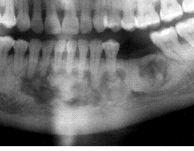
Florid cemento-osseous dysplasia. Section of a panoramic tomogram showing the typical appearances of multiple irregular radiopaque masses centred on the roots of the teeth. The periphery of each is radiolucent, and the surrounding bone shows some sclerosis. Similar lesions were present in the contralateral molar region and on some maxillary teeth

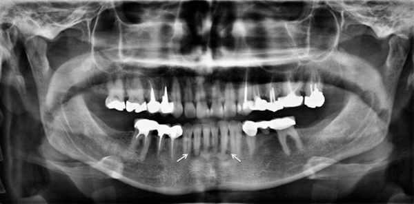
A panoramic radiograph shows a radiolucent rim at the periphery of lesions (arrows) diagnosed as periapical cemento-osseous dysplasia. Copyright © 2015 by Korean Academy of Oral and Maxillofacial Radiology
Focal cemento-osseous dysplasia
This term is given to changes similar to florid osseous dysplasia but forming a single lesion on one tooth. The lesion resembles a cemento-osseous fibroma radiographically, but shows the maturation sequence typical of this group of lesions. Lesions are usually in the posterior mandible.
Management of cemento-osseous dysplasias
Osteomyelitis starting in these lesions following extraction must be avoided because the widespread sclerosis of the late stages makes the infection difficult to treat. Wide excision may then be required to allow resolution. Treatment is otherwise not indicated except, rarely, for cosmetic reasons if there is expansion or if solitary bone cysts develop.

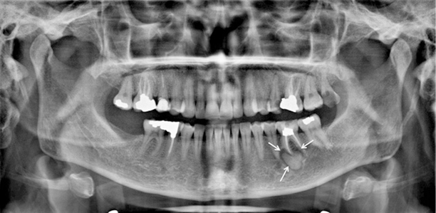
Focal cemento-osseous dysplasia is seen at the periapex of the left mandibular first molar with a radiolucent rim (arrow) on a panoramic radiographCopyright © 2015 by Korean Academy of Oral and Maxillofacial Radiology

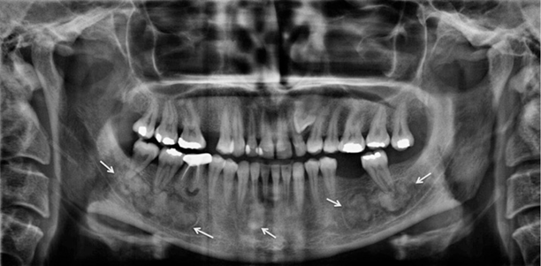
Florid cemento-osseous dysplasia is situated at the right, left, and anterior portions of the mandible, surrounded by a radiolucent rim (arrows).Copyright © 2015 by Korean Academy of Oral and Maxillofacial Radiology